カレントテラピー 36-6 サンプル page 12/34

このページは カレントテラピー 36-6 サンプル の電子ブックに掲載されている12ページの概要です。
秒後に電子ブックの対象ページへ移動します。
「電子ブックを開く」をクリックすると今すぐ対象ページへ移動します。
概要:
カレントテラピー 36-6 サンプル
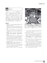
Current Therapy 2018 Vol.36 No.6 15膵腫瘍性疾患517Ⅵ おわりに膵癌のダイナミックCTによる画像診断について解説した.膵癌が疑われる症例では,高濃度,高用量のヨード造影剤を使用した多相のダイナミックCTが推奨される.膵癌は乏血性であるが,遅延性に濃染することも理解しておく必要がある.膵癌の進展度診断を行う場合には多方向から観察し,漿膜浸潤,後腹膜浸潤,神経叢浸潤,脈管浸潤,遠隔転移の有無を詳細に観察することが重要である.参考文献1)福島敬宣,向井 清:膵癌の病理像と病理学的評価.癌と化学療法 32:599-604, 20052)Fishman EK, Horton KM, Urban BA:Multidetector CTangiography in the evaluation of pancreatic carcinoma:preliminaryobservations. J Comput Assist Tomogr 24:849-853,20003)Yanaga Y, Awai K, Nakayama Y, et al:Pancreas:patientbody weight tailored contrast material injection protocol versusfixed dose protocol at dynamic CT. Radiology 245:475-482, 20074)蒲田敏文,服部由紀,望月健太郎ほか:膵管癌の画像診断―発生部位による臨床的・画像的特徴―.画像診断 30:1272-1286, 20105)Ishigami K, Yoshimitsu K, Irie H, et al:Diagnostic value ofthe delayed phase image for iso-attenuating pancreatic carcinomasin the pancreatic parenchymal phase on multidetectorcomputed tomography. Eur J Radiol 69:139-146, 20096)Brugel M, Link TM, Rummeny EJ, et al:Assessment of vascularinvasion in pancreatic head cancer with multislice spiralCT:value of multiplanar reconstructions. Eur Radiol14:1188-1195, 20047)蒲田敏文,龍 泰治,南 哲弥ほか:膵癌におけるDynamicCT撮像方法と脈管浸潤.胆と膵 32:585-592, 20118)Lu DS, Reber HA, Krasny RM, et al:Local staging of pancreaticcancer:criteria for unresectability of major vesselsas revealed by pancreatic -phase, thin -section helical CT.AJR Am J Roentgenol 168:1439-1443, 19979)Kayahara M, Nagakawa T, Konishi I, et al:Clinicopathologicalstudy of pancreatic carcinoma with particular referenceto the invasion of the extrapancreatic neural plexus . Int JPancreatol 10:105-111, 199110)Mochizuki K, Gabata T, Kozaka K, et al:MDCT findings ofextrapancreatic nerve plexus invasion by pancreas head carcinoma:correlation with en bloc pathological specimens anddiagnostic accuracy. Eur Radiol 20:1757-1767, 201011)望月健太郎,蒲田敏文,小坂一斗ほか:膵管癌の進展度診断―CTと病理組織の対比から―.画像診断 30:1287- 1297,201012)Nawaz H, Fan CY, Kloke J, et al:Performance characteristicsof endoscopic ultrasound in the staging of pancreaticcancer:a meta-analysis. JOP 14:484-497, 201313)Gabata T, Matsui O, Terayama N, et al:Imaging diagnosisof hepatic metastases of pancreatic carcinomas:significanceof transient wedge-shaped contrast enhancement mimickingarterioportal shunt. Abdom Imaging 33:437-443, 200814)日本膵臓学会:膵癌取扱い規約第7版.金原出版,東京,201615)Soloff EV, Zaheer A, Meier J, et al:Staging of pancreaticcancer:resectable, borderline resectable, and unresectablediseases. Abdom Radiol 43:301-313, 2018SMVSMAT図14 膵鉤部癌,上腸間膜動脈浸潤,上腸間膜静脈浸潤(UR)ダイナミックCT動脈相では,膵鉤部に乏血性腫瘍(T)を認める.腫瘍は前方に進展し上腸間膜動脈(SMA)および上腸間膜静脈(SMV)を巻き込んでいる.切除不能(UR)である.